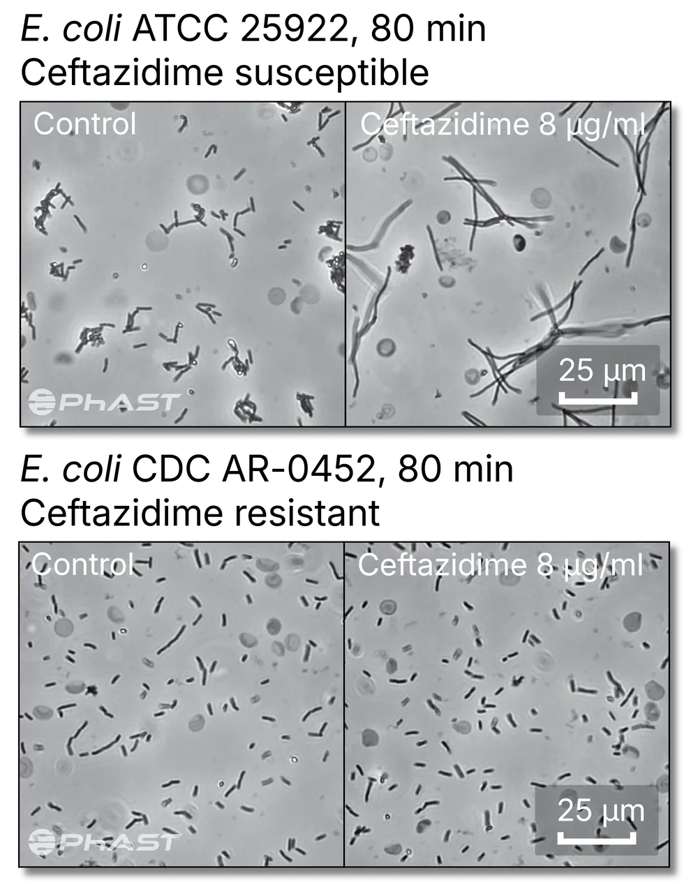
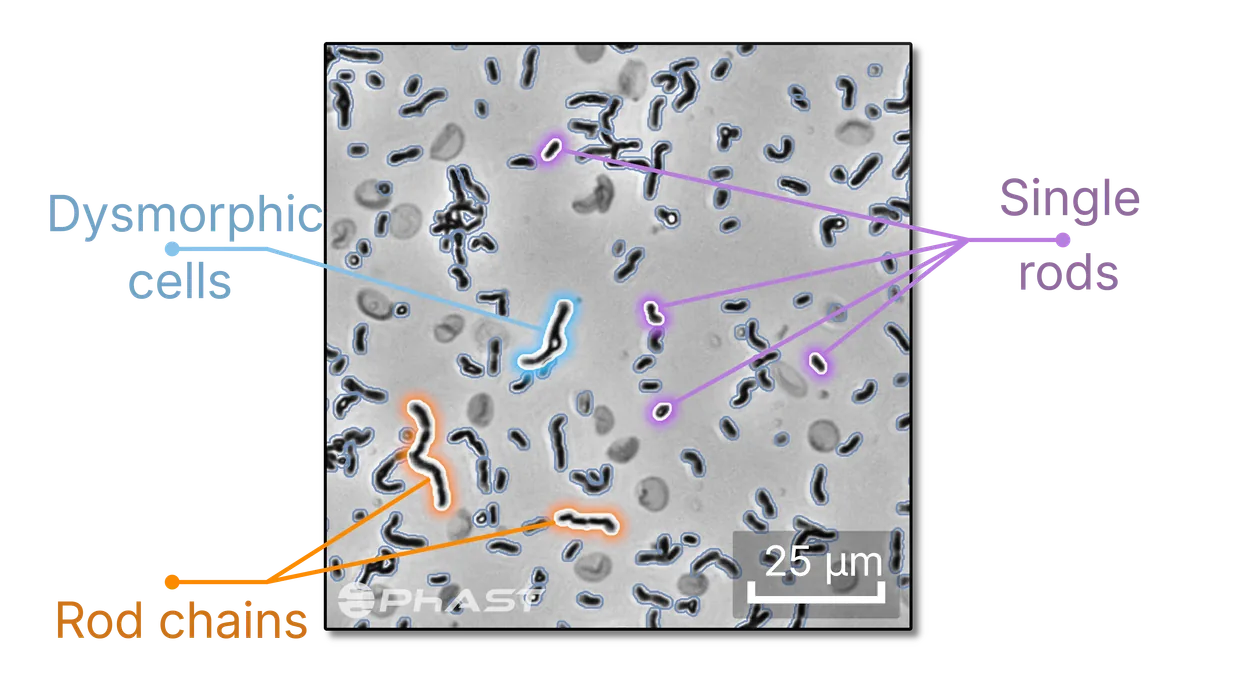
PhAST’s internally developed computer vision resolves individual bacteria among other cells and debris, with no isolation and no staining required. The analysis works from distributions of measurements taken across many individual cells, exposing details of the early stages of antimicrobial interaction. A locked machine learning pipeline converts the complex cellular response to antimicrobials into a categorical output.
PhAST builds with interpretability as a priority at all stages. It is what allows us to develop robust assays: to trace the cause of an error, and to recognize a sample outside the range the analysis was trained on.
PhAST believes this approach is a foundation for diagnostics well beyond our first assay: computer vision strong enough to characterize the form and behavior of cells in conditions close to their original specimen.
Single-cell level analysis
Early phenotypic response